CURRENT BIOMEDICAL RESEARCH IN THE BOWSER LAB
In the human kidney, fluid destined to become urine flows across the upper (lumenal) surfaces of the cells that form the renal tubules. As shown above, the primary cilium extends from the cell surface into this urinary flow field. The primary cilium is therefore ideally positioned to monitor the composition and flow rate of the nascent urine. Our latest results show that it is also a potential site for the binding of particles suspended in the renal fluid, such as those involved in kidney stone formation (i.e., calcium oxalate microcrystals). However, it has yet to be shown that primary cilia play any role in kidney physiology or pathology. Our biomedical research program is designed to study this question.
Schartz, E.A., Leonard, M.L., Bizios, R., and Bowser, S.S. (1997) Analysis and modeling of the primary cilium bending
response to fluid shear. American Jounal of Physiology 272 (Renal Physiology 41): F132-F138.
Wheatley, D.N. (1997) "Essential" function(s) of the centriole: Questions. Cell Biology International 21:191-192.
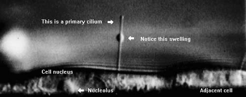
Seen here is a videomicroscopic side-view of a cultured kidney epithelial cell. Notice the long primary cilium projecting from the apical cell surface. Most kidney epithelial cells express primary cilia, as do many other human cell types. Although primary cilia are nonmotile (i.e., they do not beat), surface-attached particles and ciliary swellings are transported along the cilium shaft. The mechanism and significance of this ciliary transport are poorly understood.